‘Equine Dysautonomia’ or the disease commonly known as Equine Grass sickness (EGS), has been recognised for more than a century and most horse owners have heard or had some experience of this devastating disease.
It is (mainly) a disease of grazing horses that appears to have a seasonal and regional occurrence. Most cases of EGS are fatal, with only some chronic cases surviving with appropriate nursing care.
EGS is one of a group of diseases known as ‘dysautonomias’ (meaning gut dysfunction), that occur in a number of other species (eg cats, dogs, sheep).
The clinical signs in all species are similar and the pathology seen is very specific, making a common cause highly likely.
Equine grass sickness is also seen in a number of other countries, eg ‘Mal Seco’ – a wasting disease of horses reported in Argentina, which appears to be the same disease; therefore EGS is not only a disease seen exclusively in the United Kingdom.
The clinical signs of EGS largely reflect disruption of the nerve supply to the gastrointestinal tract, which is responsible for normal gut motility and production of faeces.
The most common presentation of EGS to veterinary surgeons is ,therefore, usually colic.
The pathology seen in EGS (and dysautonomias in other species) cases is a very specific pattern of neurodegeneration resulting in permanent loss of neurons throughout the gut.
However, the pathology of EGS is much more widespread and this is evident in the range of clinical signs seen and at post mortem examination.
Clinical signs
EGS is seen in a number of different forms: acute, subacute and chronic.
However, it is important to emphasise that these are all manifestations of the same disease and rather than distinct categories, they are more of a continuum of the disease.
The clinical signs seen with the different forms generally reflect the extent of neuronal loss/severity of the disease and the time course associated with development of clinical signs.
Acute cases are very sudden onset (usually within 24 hours), subacute cases (several days duration) and chronic cases can extend over weeks or months.
The different categories are important from a veterinary perspective, since they help to determine which horses are candidates for treatment.
Only chronic cases have some chance of survival and even within these, it is important that horses maintain some ability to eat/swallow and have sufficient gut motility to survive.
The amount of care, time, effort and emotional investment in nursing grass sickness cases should also not be underestimated.
Some of the most common signs observed in EGS cases are:- colic (of varying severity), dullness/depression, muscle tremors, patchy sweating, dysphagia (difficulty in swallowing), reduced or complete loss of appetite and ptosis (droopy eyelids or a sleepy appearance).
Horses often have a markedly increased heart rate but often appear very dull and gut sounds are also dramatically reduced or absent with no or only scant amounts of mucoid covered faeces produced.
These characteristic signs are usually more obvious and pronounced in acute and subacute cases; whereas chronic cases may show much milder or minimal signs initially and may even present with more uncommon signs such as choke and excess salivation.
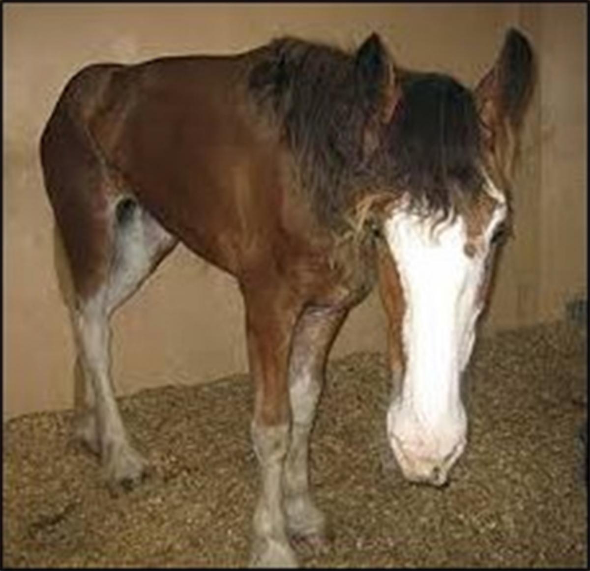
The chronic forms of EGS may present primarily as dramatic weight loss and a diagnosis of EGS may not be immediately obvious.
Horses with chronic EGS often develop a classic tucked up “grey-hound” type appearance (figure 1) and rhinitis sicca (painful sloughing and crusting of the nasal passages), which reduces their sense of smell and appetite further.
Diagnosis
The diagnosis of EGS in the live animal is based largely on the presence of classic clinical signs. A definitive diagnosis of EGS requires histopathology on a small intestinal biopsy sample and the specific pathological findings.
One of the most consistent signs of EGS is ‘ptosis’ or a sleepy appearance, with droopy upper eyelids and eyelashes.
This is due to a loss of nerve supply to the smooth muscle of the upper eyelids.
Without this nerve supply, a chemical transmitter – adrenaline cannot be delivered and the upper eyelid is unable to contract and remain open. Application of a drug phenylephrine (similar effects to adrenaline) directly to the eye can reverse the ‘ptosis’ seen in EGS cases. Therefore, phenylephrine eye drops applied to one eye, can be used to aid diagnosis of EGS in suspected cases.
Risk factors for EGS
Previous studies have helped to identify a number of risk factors associated with the development of EGS. Certain management practices have been associated with an increased risk of EGS development, for example harrowing or use of machinery, which may result in pasture or soil disturbances and potentially increase exposure to a causative agent.
The seasonal incidence of EGS (late spring/early summer) is likely to be associated with a particular set of weather conditions that support the growth of a causative agent on particular pastures (eg namely cold nights and clear bright, sunny days).
EGS cases frequently follow heavy periods of rainfall, especially if preceded by a hot, dry spell. It is possible that heavy rainfall might stir up the pasture and distribute a causative agent present, increasing exposure in grazing horses.
Age has been identified as a risk factor for developing EGS, with more cases seen in younger (2-7 years) compared to older horses. However, any new arrivals to a premises where EGS cases have previously been reported, also appear to be at increased risk of developing the disease.
It may be that immune status (age associated) and also an individual horse effect have an influence on disease susceptibility. Stress itself and stressful events (such as weaning, castration, worming, separation from pasture mates) may also affect immunity and play a role in disease susceptibility (particularly in high risk individuals).
Aetiology – what causes EGS?
Numerous studies investigating the cause of EGS have been performed over the years and these have contributed a lot to understanding more about EGS and in particular, identifying risk factors.
Recent research has focused on the bacterium Clostridium botulinum as the most likely cause of EGS; with some sort of dietary trigger causing overgrowth of this bacteria and toxin production inside the horses gut. This was based on observations that EGS cases had increased levels of this bacteria inside their gut. Co-grazing horses (unaffected horses grazing in the same field as EGS cases), also had increased levels of Clostridium botulinum antibodies in their bloodstream and surviving chronic EGS cases had higher antibody levels compared to non-surviving cases.
These findings suggest that having antibodies against Clostridium botulinum may offer some protection against developing EGS.
They also formed the basis of a large scale Clostridium botulinum vaccination trial in horses on previously affected premises.
The results of this trial have yet to be fully disclosed; but could offer some hope in the prevention of EGS in the future.
More recently, a number of observations in respect to EGS have led to the re-consideration of a fungal aetiology.
For example, the clinical signs of EGS cases are very different to any of the other Clostridium associated diseases. Many of the identified risk factors for EGS are consistent with involvement of a fungus; including the strong association with grazing, particular soil types, seasonal occurrence, clustering of cases within certain geographical locations and certain weather patterns that could support fungal growth.
A more recent and significant contribution to support a fungal cause would also be the very specific pathology seen in EGS (and not botulism) cases.
Current research being undertaken at the University of Edinburgh, R(D)SVS and Roslin Institute is investigating the potential role of fungal toxins (‘mycotoxins’) in EGS. It is proposed that EGS could be caused by a neurotoxin produced by a fungus growing on the pasture and ingested by grazing horses. Earlier studies investigating a fungus and EGS have been inconclusive.
However, these investigations were limited by the inability to isolate and identify many of the fungi present in the equine gastrointestinal tract using standard laboratory techniques.
Advances in technology have since revolutionised the ability to detect agents or toxins using a gene sequencing approach. These are the methods being used in this current approach. The results of this research are close to completion and it is hoped that the information gained will provide further insight into preventing EGS in the future.
Prevention
Currently, any recommendations for the prevention of EGS are based on sensible use of the information gained from previous epidemiological studies. Stressful events (eg weaning, castration, separation from pasture mates) should be avoided (if possible) during peak periods of EGS (late spring, early summer months).
This, however, is becoming increasingly difficult with highly changeable weather patterns and recognising particular weather conditions that appear to precede the onset of EGS cases could be more helpful (cold nights; bright, sunny days).
Horses should be provided with supplementary forage, particularly on heavily grazed pastures, as this may discourage rummaging, less pasture disruption and decreased exposure to a causal agent.
Faeces should ideally be removed from the pasture as this may reduce exposure to a causative agent.
However, droppings should be removed manually rather than using mechanical devices. In addition, some pasture management practices (eg harrowing) may disrupt and again increase exposure to an agent on the pasture.
The use of pastures immediately following any potential pasture disruption should also be avoided. Ideally and if possible, any pastures previously associated with EGS cases should not be grazed by high risk animals (younger animals, new arrivals) during peak EGS season.
The information provided within this review is an overview of equine grass sickness and is attributable to colleagues at the R(D)SVS, Roslin and Moredun Institutes. More extensive information can be found at www.grassickness.org.uk


Comments: Our rules
We want our comments to be a lively and valuable part of our community - a place where readers can debate and engage with the most important local issues. The ability to comment on our stories is a privilege, not a right, however, and that privilege may be withdrawn if it is abused or misused.
Please report any comments that break our rules.
Read the rules hereComments are closed on this article