
Johne's disease is found in all sheep husbandry systems including extensively managed flocks and is characterised by emaciation but not, as in cattle, chronic severe diarrhoea.
In infected flocks, ewe mortality rate from Johne's is estimated to be as high as 5-10% per year.
The causal bacterium, Mycobacterium avium subspecies paratuberculosis, survives on pasture for many months, with infected sheep developing clinical disease via the faecal/oral route. Infection can also be acquired in utero during the advanced stages of disease in the ewe.
However, infection ingested by adult sheep is unlikely to cause clinical disease. Goats can be an important source of infection when co-grazed with sheep. The role of cattle and various wildlife species including rabbits, in the epidemiology of Johne's disease, is currently under investigation.
Clinical presentation
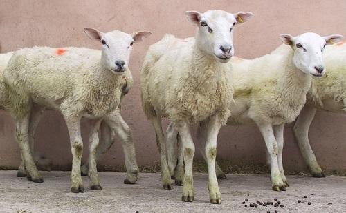
Sheep with Johne's disease show chronic weight loss/low body condition score with a poor fleece in individual middle-aged (typically three to four years) sheep even when fed an appropriate plane of nutrition.
Emaciated sheep are typically detected during routine flock handling procedures, such as pre-mating checks, when their body condition score of 1.5 or below (scale 1 to 5) against others managed in a similar way with scores of 3.0 or greater.
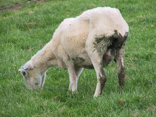
Chronic diarrhoea, unlike cattle, is not a feature in the majority of sheep affected by paratuberculosis and sheep often void pelleted faeces, unless there is also a heavy concurrent parasitic burden. Affected sheep appear bright and alert with a normal appetite but rumen fill is reduced.
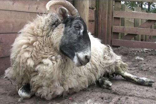
Wool is poor quality and the fleece appears more open than usual. During the final stages affected sheep may become recumbent and too weak to stand. Submandibular oedema (bottle jaw) presents only in extreme cases. The severity of Johne's disease is compounded by concurrent parasitism, especially fasciolosis.
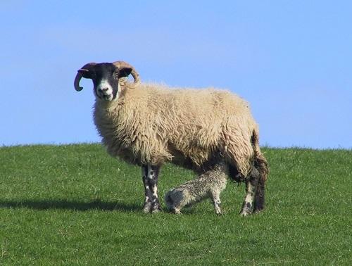
Lambs born to ewes in the terminal stages of Johne's disease have low birthweights (often as low as 2-3kg), due to chronic intra-uterine growth retardation
Differential diagnoses
In order of decreasing importance, the common causes of weight loss are:
Group problem:
Poor flock nutrition
Fasciolosis
Chronic parasitism due to poor pasture management and erroneous control strategies
Chronic parasitism caused by anthelmintic-resistant strains of nematodes
Virulent footrot
Individual sheep:
Poor dentition especially molar teeth
Chronic pneumonia, mastitis, or other septic focus
Chronic severe lameness
Ovine pulmonary adenocarcinoma
Intestinal adenocarcinoma
Diagnosis
The intestinal changes caused by Johne's disease cannot reliably be diagnosed by an on-farm post mortem even when undertaken by experts, with a single post mortem providing little information regarding the prevalence of certain disease conditions.
In most situations it will prove more useful to investigate weight loss in 10 or more ewes than to send a single ewe for detailed post mortem examination. Serum albumin and globulin determinations provide your veterinary surgeon with the most useful screening tests for the investigation of chronic weight loss in a number of adult sheep.
Sheep with Johne's disease have profound hypoalbuminaemia and a normal globulin concentration. These changes result from loss of albumin across the damaged gut.
It must be remembered that sheep with Johne's co-incidentally often have very high parasitic faecal egg counts indeed and such results may lead to erroneous conclusions. Sheep with Johne's disease also cause considerable pasture contamination due to this very high egg production.
Significant serological titres (either AGID or ELISA) are only detected in approximately 60% of clinical paratuberculosis cases resulting in a high false negative rate (low sensitivity) although the specificity is high (greater than 95%).
Furthermore, direct faecal examination provides unreliable results as there may be few bacteria present.
Cultural isolation taking at least eight weeks, and possibly up to six months, has very limited practical application.
Post mortem
There is an emaciated carcase with gelatinous atrophy of fat depots. Thickening of the ileum with prominent ridging is not always obvious in Johne's disease but the mesenteric lymph nodes are often visibly enlarged. The diagnosis is confirmed after ZN staining of ileal sections and ileo-caecal lymph nodes which demonstrate clumps of acid-fast bacteria.
Management/Prevention/Control measures
The true Johne's disease incidence in UK sheep flocks remains unknown but many flocks have losses in excess of 5% without appreciating the cause of such losses.
Encouraging results with much reduced disease prevalence have been reported following adoption of a vaccination programme in several countries.
Vaccination offers the best long-term prospect for control of Johne's disease in sheep flocks (approximately £2-3 per dose).
Economics
Difficulties with confirmation of the provisional diagnosis of Johne's disease, coupled with very few ewes culled for poor condition/emaciation submitted to the veterinary surgeon, lead to a gross underestimation of the prevalence and financial impact of this disease in the UK.
* This article was provided by the National Animal Disease Information Service


Comments: Our rules
We want our comments to be a lively and valuable part of our community - a place where readers can debate and engage with the most important local issues. The ability to comment on our stories is a privilege, not a right, however, and that privilege may be withdrawn if it is abused or misused.
Please report any comments that break our rules.
Read the rules here