

Osteochondrosis Dissecans and Bone Cysts in the horse
Osteochondrosis Dissecans (OCD) is a relatively common developmental orthopaedic disease in horses. It can occur in all breeds and causes lameness in 5-25% of affected horses. In general larger horses are more commonly affected than ponies.
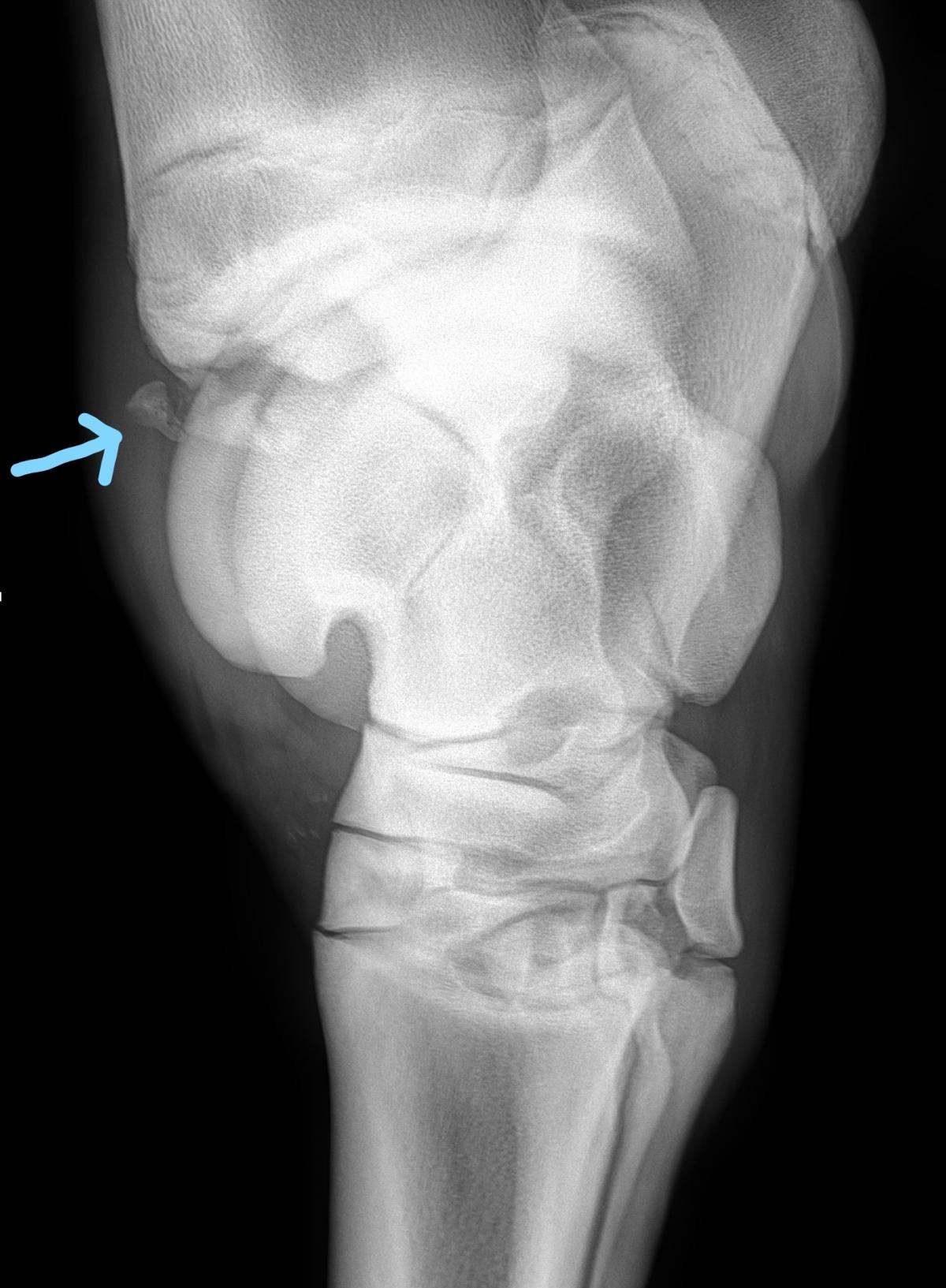
In a healthy joint the bone is covered in a layer of articular (or joint) cartilage, allowing the smooth movement of the joint articulation. In a joint with OCD the cartilage develops as a foal with variable thickness and strength. As a result, the cartilage is weaker than it should be which results in flaps of the cartilage and bone breaking off from the parent bone later in life. This usually occurs in the gliding areas of the joint and the loose fragments do not re-adhere to the bone but remain partially attached and loose. Occasionally the fragments break away entirely and float around in the joint. The loose or floating fragments of bone cause inflammation and irritation to the synovial lining of the joint resulting in the clinical signs of joint inflammation.
The cause of the disease process is not abundantly clear, although it is thought to be multifactorial. Known predisposing factors include excess or imbalances in nutrition, (in particular carbohydrates and mineral imbalances), joint trauma and excess exercise, endocrine (hormonal) factors and in some cases genetic predisposition.
Clinical signs
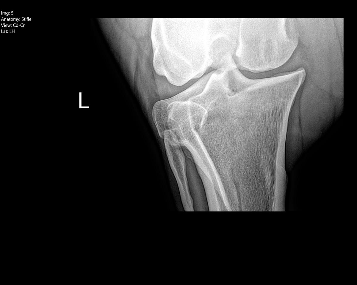
Horses with OCD typically initially present with clinical symptoms between 4 months and 2 years of age, although they can present later in life. Most cases present with swelling of the affected joint, or joints, and up to 25% of them present with lameness (often lameness is not obvious at walk but evident at trot or canter). The lameness is variable depending on the joint location and severity of the lesion. The most commonly affected joints are the hocks, stifles and fetlocks, although lesions can occur in any joint.
Diagnosis
Diagnosis of OCD is made with a thorough clinical examination, regional anaesthesia of the limb and x-rays of the affected joint. Generally, we x-ray paired joints (each leg) to ensure that it isn’t a bilateral problem. Occasionally lesions can be identified on x-rays of older horses, presenting for different clinical reasons, and are not of any clinical concern. Ultrasound can also be a helpful imaging modality to determine the extent of the lesion, this is case dependent.
Treatment and Prevention
Treatment options for OCD depends on the size of the fragment, the site within the joint and the joint in question. Some joints, for example the shoulder, have a poor prognosis for treatment whatever option is taken. Other joints have a much better prognosis and a more conservative treatment regime can be taken
For the majority of cases (for the average hock and stifle OCD for example) the preferred treatment of choice is general anaesthesia and key-hole surgery (arthroscopy) to remove the fragments at a young age. This is a minimally invasive technique with good success rate. The joint is also lavaged with a large volume of sterile fluid to remove the joint inflammatory markers at the time of the surgery. The key-hole portals are stitched closed, these are removed 10-14 days after the surgery. The horse is normally hospitalized for 2-3 days following the surgery to ensure there are no post- operative complications. Depending on the affected joint, a bandage will be in place, usually until stitches are removed. A case dependent rehabilitation programme will be given for the following weeks and months.
In cases where keyhole surgery is not performed the horse will on average have a reduced prognosis for athletic soundness. The presence of the fragment can result in the joint becoming arthritic as the horse ages.
Prevention starts from a foal, ensuring the correct diet; do not feed excessive carbohydrates, this can cause rapid growth which has been shown to be a contributing factor to the development of OCD. It should also be ensured that the correct balance of vitamins and minerals are being provided.
It is also imperative that exercise of the foal is controlled in the early months to prevent excessive forces and trauma being applied to the joint.
Bone Cysts
Bone cysts is another form of developmental bone disease that also results due to poor or weak cartilage formation. It tends to occur at the weight bearing areas of a joint. With this weak cartilage and underlying bone, the area collapses to form a defect into which joint fluid may gain access. By far the most common site for the formation of a bone cyst is in the stifle joint although all other joints can be affected, particularly the pastern and coffin joints.
The signs develop at a similar age to OCD and the horses are usually lame with joint distension.
Treatment of bone cysts is complex and is based on a case by case basis. It has also changed over the years. The current treatment of choice is to inject steroids directly into the cyst or to place a screw across the cyst to provide a stable platform to support the collapsed and imploded cartilage and bone.


Comments: Our rules
We want our comments to be a lively and valuable part of our community - a place where readers can debate and engage with the most important local issues. The ability to comment on our stories is a privilege, not a right, however, and that privilege may be withdrawn if it is abused or misused.
Please report any comments that break our rules.
Read the rules here